The Problem: Fibrosis Is the Silent Killer After Heart Attack
Every year, approximately 17 million people worldwide suffer an acute myocardial infarction (MI). Thanks to advances in emergency cardiology — thrombolysis, percutaneous coronary intervention (PCI), and improved critical care — more patients than ever survive the initial event. But survival carries a hidden cost.
The Post-MI Remodeling Cascade
When a heart attack kills a portion of the myocardium, the body launches a wound-healing response. Immune cells flood the injured area to clear dead tissue, and fibroblasts deposit collagen to form a scar — a process called fibrosis. In the short term, this scar holds the damaged heart together. But in the weeks and months that follow, the fibrotic response often overshoots: excessive collagen deposition stiffens the ventricular wall, distorts its geometry, and impairs the heart’s ability to pump effectively. This process, known as adverse ventricular remodeling, is the primary driver of post-MI heart failure.
Current therapies — ACE inhibitors, beta-blockers, mineralocorticoid receptor antagonists — can slow remodeling, but they cannot reverse the fibrosis that has already formed. Once collagen has replaced functional myocardium, no existing drug can convert scar tissue back into contractile muscle or even meaningfully reduce its extent. The fibrosis is, in clinical terms, irreversible.
The Numbers Behind the Burden
Heart failure following MI carries a five-year mortality rate of approximately 50% — comparable to many cancers. In China alone, an estimated 8.9 million people live with heart failure, and post-MI fibrosis is the leading cause. The global burden is measured in tens of millions of patients and hundreds of billions of dollars in healthcare costs. Any intervention that could reverse fibrosis — not merely slow it — would represent a paradigm shift in cardiovascular medicine.
What Are iDCs? Engineering an Immune Peacekeeper
Dendritic cells are traditionally known as the sentinels of the immune system — antigen-presenting cells that activate T cells and launch immune responses against pathogens and tumors. But dendritic cells are remarkably plastic. Under the right conditions, they can be reprogrammed to become tolerogenic — suppressing immune responses rather than activating them.
The Engineering Process
The SAHZU team developed a method to derive immunosuppressive conventional dendritic cells (iCDCs) from bone marrow precursors. The process involves:
- Isolation: Harvesting bone marrow-derived mononuclear cells from the patient or donor
- Directed differentiation: Culturing these precursors with a specific combination of cytokines and growth factors that steer them toward the conventional dendritic cell lineage rather than the plasmacytoid dendritic cell lineage
- Immunosuppressive conditioning: Treating the differentiating cells with immunomodulatory agents — including rapamycin (an mTOR inhibitor) and IL-10 — that reprogram their functional phenotype from immune-activating to immune-suppressing
- Verification: Confirming surface marker expression consistent with tolerogenic dendritic cells (high PD-L1, high ILT3/ILT4, low co-stimulatory molecules CD80/CD86) and functional suppressive capacity in vitro
The result is a cell that, when introduced into the fibrotic heart, acts as an immune peacekeeper — dampening the chronic inflammation that drives ongoing fibrosis and, remarkably, signaling existing fibroblasts and macrophages to shift from a pro-fibrotic to a pro-resolution phenotype.
Why Dendritic Cells, Not Tregs?
Regulatory T cells (Tregs) have been the focus of many immune-based approaches to fibrosis, but they face a critical limitation: Tregs are difficult to expand ex vivo to therapeutic quantities, they have limited persistence after infusion, and their suppressive function is highly context-dependent. Dendritic cells, by contrast, are natural orchestrators of the immune environment — they do not merely suppress, they actively reprogram the tissue microenvironment. A single dendritic cell can modulate dozens of T cells, macrophages, and other immune effectors, making them far more efficient modulators on a per-cell basis.
The Nature Study: Design and Results
The landmark study, published in Nature on April 8, 2026 (DOI: 10.1038/s41586-026-10346-5), was led by Xinyang Hu and Yang Xu from the Second Affiliated Hospital of Zhejiang University School of Medicine (SAHZU), with collaborators from multiple Chinese and international institutions.

Murine Model: Proof of Concept
The team first established proof of concept in a mouse model of myocardial infarction. Mice underwent permanent left anterior descending (LAD) coronary artery ligation, producing a well-characterized model of post-MI fibrosis. iCDCs were administered via intravenous injection at defined time points after infarction.
The results were striking:
- Fibrosis reduction: iCDC-treated mice showed a 40-60% reduction in cardiac fibrosis area compared to controls, as measured by Masson’s trichrome staining and picrosirius red polarization
- Ejection fraction improvement: Cardiac MRI and echocardiography demonstrated significant preservation and partial recovery of left ventricular ejection fraction
- Survival benefit: iCDC-treated mice had significantly improved long-term survival compared to vehicle-treated controls
- Immune microenvironment shift: Flow cytometry and single-cell RNA sequencing revealed a dramatic shift in the cardiac immune landscape — pro-fibrotic M2-like macrophages decreased, pro-resolution macrophages increased, and pathogenic fibroblast populations contracted
Mechanistic Insights from Single-Cell Analysis
Using single-cell RNA sequencing (scRNA-seq) of cardiac tissue from treated and untreated animals, the team mapped the transcriptional changes induced by iDC therapy. Key findings included:
- Fibroblast reprogramming: Pro-fibrotic fibroblasts (expressing high levels of collagen type I, type III, and periostin) downregulated their extracellular matrix production program and adopted a more quiescent or even pro-resolution phenotype
- Macrophage polarization shift: The balance of cardiac macrophage populations shifted from pro-fibrotic (TGF-beta-producing, Arg1-high) to pro-resolution (IL-10-producing, CD163-high) phenotypes
- T cell modulation: CD4+ T cells in the iDC-treated hearts showed increased expression of anti-inflammatory markers and decreased production of pro-fibrotic cytokines
The Primate Breakthrough: From Mice to Macaques
Mouse models are essential for proof of concept, but the translational gap between rodent and human cardiology is well documented. The cardiovascular system of a mouse — with a heart rate of 500-600 beats per minute and a cardiac physiology adapted to a creature weighing 25 grams — differs fundamentally from that of a human. To bridge this gap, the SAHZU team conducted the critical next step: validation in non-human primates.

The Cynomolgus Macaque Study
The team used cynomolgus macaques (Macaca fascicularis), whose cardiovascular physiology closely mirrors that of humans — similar heart rates, similar coronary anatomy, and similar post-infarction remodeling patterns. Macaques underwent experimentally induced myocardial infarction via temporary coronary artery occlusion, followed by reperfusion — a model that closely replicates the clinical scenario of a human patient who receives timely PCI.
iCDCs derived from each animal’s own bone marrow (autologous iDCs) were infused intravenously after infarction. The results were nothing short of transformative:
- Fibrosis reversal: Not only was further fibrosis halted, but existing fibrotic tissue was measurably reduced — a finding never before achieved with any cell therapy in a primate infarction model
- Cardiac function recovery: Ejection fraction, which typically declines after MI and remains depressed, showed meaningful improvement in iCDC-treated animals
- Left ventricular remodeling attenuation: The progressive dilation and thinning of the ventricular wall that characterizes adverse remodeling was significantly reduced
- Safety profile: No adverse events, arrhythmias, or off-target organ effects were observed — a critical finding for any cell therapy approaching clinical trials
Why Primate Validation Is the Decisive Step
The failure rate for therapies that succeed in mice but fail in humans is estimated at over 90% in cardiovascular medicine. The primate model dramatically narrows this translational gap because primate hearts share with human hearts: similar coronary artery anatomy (including the prevalence of a left-dominant circulation), similar post-infarction inflammatory dynamics, similar fibroblast biology, and similar immune cell populations. By demonstrating fibrosis reversal in primates, the SAHZU team has cleared the most critical preclinical hurdle.
How iDCs Work: The Molecular Mechanism
Understanding how iDCs reverse fibrosis requires looking at the molecular crosstalk between these engineered cells and the cardiac tissue microenvironment. The mechanism is not a single pathway but a coordinated multi-level reprogramming of the fibrotic niche.
The PD-L1/PD-1 Axis
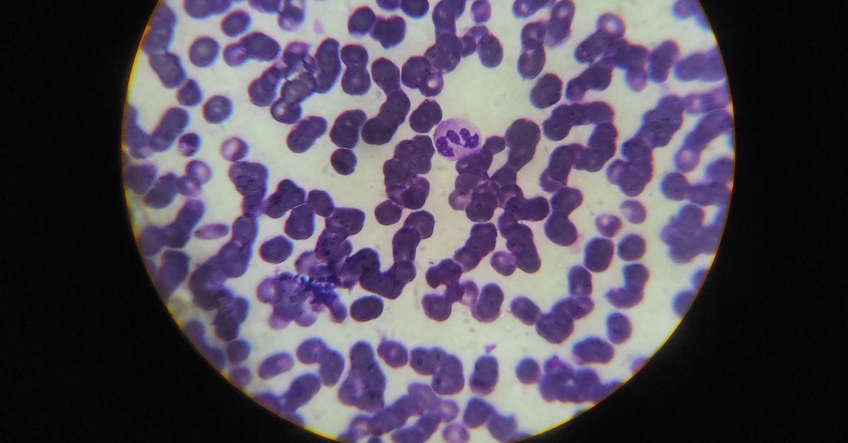
iDCs express high levels of PD-L1 (programmed death-ligand 1), which engages PD-1 on activated T cells and pro-fibrotic macrophages. This interaction delivers an inhibitory signal that dampens the pro-inflammatory and pro-fibrotic activity of these effector cells. In the context of post-MI fibrosis, where chronic immune activation drives ongoing collagen deposition, this checkpoint engagement effectively removes the signal that sustains the fibrotic response.
IL-10 and TGF-Beta Rebalancing
iDCs secrete high levels of IL-10, an anti-inflammatory cytokine that directly suppresses fibroblast collagen production and promotes macrophage polarization toward a pro-resolution phenotype. Critically, while TGF-beta is the master driver of fibrosis, its signaling is context-dependent — in the presence of appropriate immune modulation, TGF-beta can promote tissue repair rather than fibrosis. iDCs appear to shift the TGF-beta balance from its pro-fibrotic to its pro-repair function.
Metabolic Reprogramming of the Fibrotic Niche
Emerging evidence from the study suggests that iDCs also alter the metabolic environment of the fibrotic heart. Fibrotic tissue is characterized by a hypoxic, lactate-rich microenvironment that reinforces fibroblast activation and immune cell dysfunction. iDCs appear to promote oxidative metabolism in surrounding cells, breaking the metabolic feedback loop that sustains fibrosis.
Why This Matters: Clinical Implications
The clinical implications of iDC therapy extend well beyond post-MI fibrosis. If the primate results translate to human trials — and the primate data provide strong justification for optimism — iDC therapy could become the first treatment in cardiovascular medicine that actually reverses established fibrosis.
Immediate Application: Post-MI Heart Failure
The most direct application is for patients who have survived a heart attack but are developing or have developed heart failure due to post-infarction fibrosis. Current guidelines offer neurohormonal blockade to slow progression; iDC therapy could offer the possibility of regression — reducing scar burden, improving cardiac function, and potentially preventing the progression to advanced heart failure.
Broader Applications: Fibrosis Beyond the Heart
Fibrosis is not limited to the heart. It is the final common pathway of chronic injury in the liver (cirrhosis), kidney (renal fibrosis), lung (pulmonary fibrosis), and many other organs. The immune-mediated mechanism of iDC therapy — reprogramming the tissue microenvironment from pro-fibrotic to pro-resolution — is not organ-specific. If iCDCs can reverse cardiac fibrosis, the same approach may be adaptable to other fibrotic diseases, each of which currently lacks effective anti-fibrotic treatments.
Autologous Cell Therapy Safety Advantage
Because iDCs are derived from the patient’s own bone marrow, the therapy carries no risk of immune rejection, no requirement for HLA matching, and no need for immunosuppressive drugs. This is a significant advantage over allogeneic cell therapies, which carry graft-versus-host risks and require immune suppression that is particularly undesirable in cardiovascular patients.
The SAHZU Team and Institutional Context
The Second Affiliated Hospital of Zhejiang University School of Medicine (SAHZU, 浙江大学医学院附属第二医院) is one of China’s oldest and most prestigious medical institutions. Founded in 1869, it is a comprehensive tertiary hospital with a national reputation for clinical innovation, particularly in cardiology, neurosurgery, and emergency medicine.
Key Researchers
Professor Xinyang Hu is a cardiologist and immunologist at SAHZU whose research focuses on the intersection of immune regulation and cardiovascular disease. His laboratory has been a pioneer in understanding how immune cell phenotypes influence cardiac remodeling after injury.
Professor Yang Xu is an immunologist specializing in dendritic cell biology and tolerogenic immune engineering. His expertise in reprogramming dendritic cells for therapeutic purposes has been instrumental in developing the iDC platform.
Institutional Strengths
SAHZU’s cardiovascular center is among the highest-volume in China, performing thousands of PCI procedures and cardiac surgeries annually. This clinical depth, combined with the hospital’s research infrastructure — including a GMP-grade cell manufacturing facility — positions SAHZU uniquely to advance iDC therapy from preclinical validation to clinical trials.
What International Patients Should Know
While iDC therapy for cardiac fibrosis is still in the preclinical stage and not yet available as a clinical treatment, the rapid progression from murine proof of concept to primate validation suggests that clinical trials may begin within the next 2-3 years.
Current Status and Timeline
- Preclinical stage: The therapy has been validated in mice and cynomolgus macaques with robust safety and efficacy data published in Nature
- Regulatory pathway: Autologous cell therapies follow a well-established regulatory pathway in China, and SAHZU has experience with IND (Investigational New Drug) applications for cell-based therapeutics
- Projected timeline: If IND approval is obtained, Phase I/II clinical trials could begin as early as 2027-2028
For Patients With Post-MI Heart Failure
International patients with post-infarction heart failure who are interested in emerging therapies should monitor SAHZU’s clinical trial announcements. Zhejiang University’s international patient services can facilitate enquiries about trial enrollment when the time comes. Currently, standard post-MI care — including optimal medical therapy, cardiac rehabilitation, and device therapy where indicated — remains the standard of care.
Zhejiang University and Hangzhou
SAHZU is located in Hangzhou, the capital of Zhejiang Province — one of China’s most developed and international cities. Hangzhou is covered under China’s 240-hour transit visa exemption and 30-day unilateral visa-free entry policies, making it accessible for international patients from 55+ countries without requiring a traditional visa. The city is a 45-minute high-speed rail ride from Shanghai.
Sources
- Hu X, Xu Y, et al. “Engineered immunosuppressive conventional dendritic cells reverse cardiac fibrosis in non-human primates.” Nature, 8 April 2026. DOI: 10.1038/s41586-026-10346-5
- Second Affiliated Hospital of Zhejiang University School of Medicine (SAHZU) Official Website: SAHZU International
- Talman V, Ruskoaho H. “Cardiac fibrosis in myocardial infarction—from repair and remodeling to regeneration.” Cell Death & Disease, 2016;7(2):e2062. DOI: 10.1038/cddis.2016.20
- Kong P, Christia P, Frangogiannis NG. “The pathogenesis of cardiac fibrosis.” Cellular and Molecular Life Sciences, 2014;71(4):549-574. DOI: 10.1007/s00018-013-1349-6
- Frangogiannis NG. “Cardiac fibrosis: Cell biological mechanisms, molecular pathways and therapeutic opportunities.” Molecular Aspects of Medicine, 2023;92:101161. DOI: 10.1016/j.mam.2022.101161