By omuat.com | June 21, 2026
NEC: The Silent Killer of Premature Babies
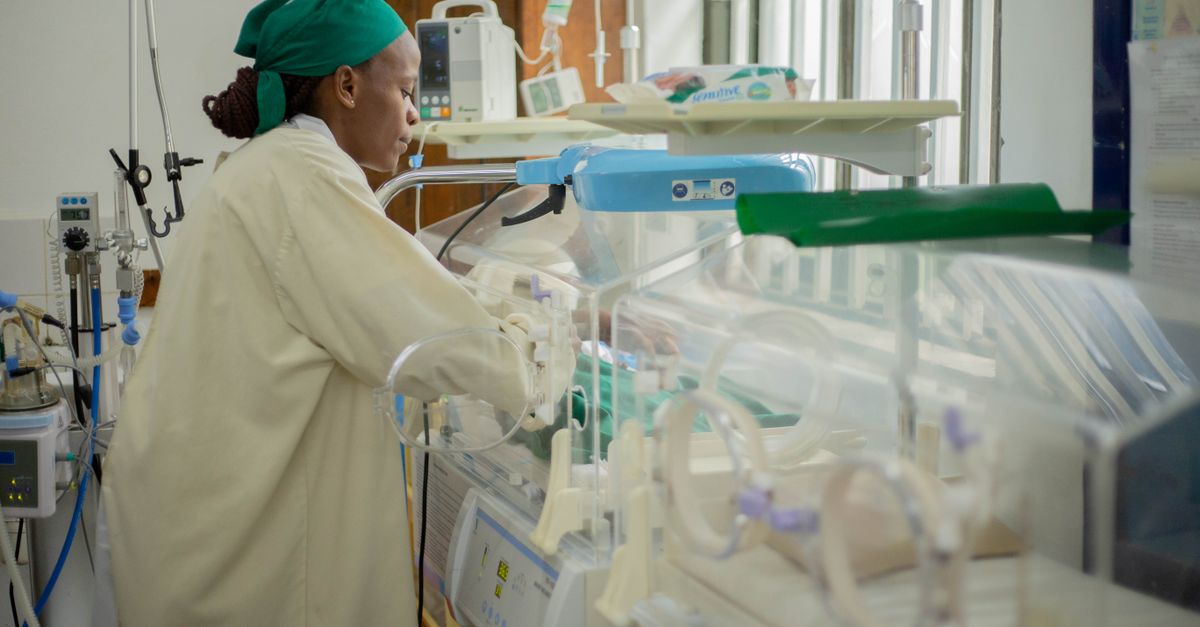
Necrotizing enterocolitis (NEC) is every neonatologist’s nightmare. It strikes without warning in premature infants — often those who seemed to be improving — and can destroy portions of the intestine within hours. With mortality rates reaching 30–50% in severe cases and survivors facing lifelong complications including short bowel syndrome and neurodevelopmental impairment, NEC remains the leading cause of death from gastrointestinal disease in premature newborns. Despite decades of research, no targeted therapy exists. Treatment remains limited to supportive care, antibiotics, and emergency surgery when the intestine perforates.
Identifying the Culprit: Double-Negative ILC3s
On May 19, 2026, a research team led by Dr. Yuxiong Guo from Guangdong Provincial People’s Hospital and Professor Yumei He from Southern Medical University published a groundbreaking study in Nature Communications that identifies — for the first time — the specific immune cell subtype responsible for driving NEC: a previously overlooked population called NKp46⁻CCR6⁻ double-negative innate lymphoid cells 3 (DN ILC3s).
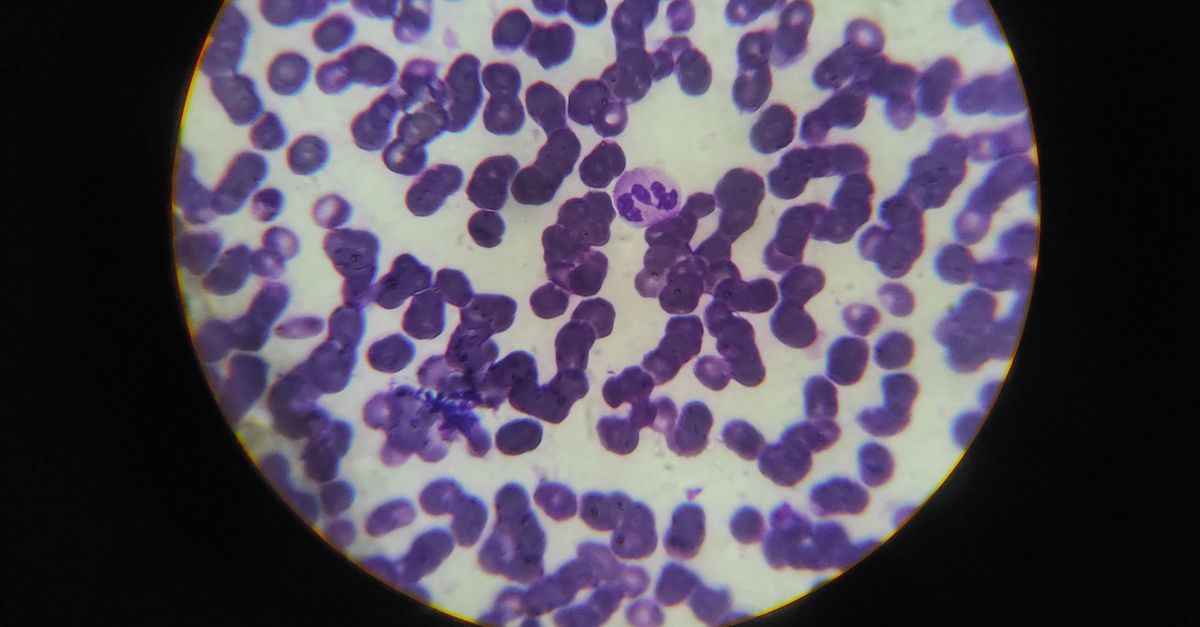
Innate lymphoid cells (ILCs) are tissue-resident immune cells that act as first responders at barrier surfaces like the intestinal lining. While ILC3s are normally protective — producing IL-22 to maintain gut integrity — the team discovered that a specific subset lacking both NKp46 and CCR6 surface markers goes rogue in NEC, secreting massive amounts of IL-17A, a potent inflammatory cytokine that directly disrupts the intestinal barrier and fuels tissue destruction.
Autophagy Gone Wrong: How Metabolism Fuels Destruction
The team’s second breakthrough was uncovering the molecular mechanism that transforms DN ILC3s from benign residents into inflammatory aggressors. The culprit is Atg5-mediated autophagy — a cellular recycling process that, in DN ILC3s, becomes hijacked to power their inflammatory metabolism.
During NEC, Atg5 activates autophagy in DN ILC3s, which in turn increases HIF-1α chromatin accessibility and transcriptional activity. This shifts the cells’ metabolism from a balanced state to aggressive glycolysis, providing the energy needed for massive IL-17A production. When the team conditionally knocked out Atg5 in RORγt⁺ cells (which include ILC3s), the results were dramatic: NEC severity was significantly reduced, DN ILC3 accumulation decreased, and IL-17A production plummeted.
A Surprising Metabolic Switch and Therapeutic Target
Atg5 deficiency didn’t just reduce inflammation — it fundamentally rewired DN ILC3 metabolism. Without autophagy, the cells shifted from glycolysis to fatty acid oxidation, a less inflammatory metabolic pathway. Using lipidomics, the team identified phosphatidylcholine as a key downstream metabolite of the Atg5-mediated autophagy pathway.
This finding is significant because it reveals that the metabolic fate of DN ILC3s — whether they become destructive or quiescent — is determined by the Atg5-autophagy-glycolipid metabolic axis. Block autophagy, and the cells revert to a less inflammatory state.

Phosphatidylcholine: A Simple Lipid That Saves Intestines
Perhaps the most clinically exciting finding was that phosphatidylcholine supplementation — a readily available dietary lipid — could suppress DN ILC3-driven inflammation, restore metabolic homeostasis, increase beneficial Clostridium bacteria abundance, and ameliorate NEC in mice. This suggests a potential therapeutic approach that is both targeted and practical: modulating the lipid environment to tame the rogue immune cells from within.
This phosphatidylcholine finding bridges immunology and the microbiome, showing that the metabolic byproducts of autophagy in immune cells directly influence gut microbial composition. The synergy between immune cell metabolism, barrier integrity, and microbial ecology creates new therapeutic intersections that had not been previously appreciated in NEC.
Human Validation and Clinical Implications
The findings extend beyond mice. Critically, human NEC tissue samples showed increased ILC3 proportions, elevated autophagic activity, and heightened IL-17A/IL-22 secretion — mirroring the mouse model findings and confirming clinical relevance.
This study, from Guangdong Provincial People’s Hospital’s PICU team led by Dr. Yuxiong Guo, is part of a remarkable series of three publications in top-tier journals (Advanced Science, Cell Death & Disease, and Nature Communications) that systematically unravel how immune cell metabolism governs neonatal and perinatal inflammatory diseases. Together, they establish a new paradigm: targeting mucosal immune cell metabolic pathways to restore tissue homeostasis is the next frontier for treating NEC and related conditions.
For families of premature infants worldwide, these findings offer genuine hope: the day may be approaching when NEC is no longer a terrifying unpredictability but a preventable and treatable condition — guided by an understanding of the rogue immune cells that drive it and the metabolic levers that can tame them.
Sources
- He J, Chen M, Peng L, et al. Blockade of NKp46⁻ CCR6⁻ ILC3 autophagy protects against necrotizing enterocolitis by restoring energy metabolism balance in mice. Nature Communications. 2026 May 19. DOI: 10.1038/s41467-026-73356-x
- Guangdong Provincial People’s Hospital. “郭予雄团队院校合作模式再获突破 三篇顶刊解锁新生儿及围产期疾病全新发病机制.” June 2, 2026. Official news release
- Chen M, et al. Probiotic intervention inhibits intestinal PMN-MDSC ferroptosis to maintain gut barrier and alleviate neonatal NEC. Cell Death & Disease. 2026. DOI: 10.1038/s41419-026-08869-w
- Chen M, et al. Trophoblast-PMN-MDSC crosstalk drives metabolic reprogramming to ameliorate advanced maternal age-related fetal growth restriction. Advanced Science. 2026. DOI: 10.1002/advs.202513370