omuat.com | June 10, 2026
 Image: Eye examination for kidney disease screening (Credit: nrd, Unsplash)
What if a simple photograph of your eye could reveal the secrets of your kidney health? Researchers at Sun Yat-sen University have developed an AI-powered system that diagnoses chronic kidney disease from retinal images - eliminating the need for invasive biopsies.
Table of Contents
- The Problem: Kidney Disease Detection Challenges
- The Breakthrough: Eye-Kidney Connection
- AI-Powered Diagnostic Technology
- Multi-Center Clinical Validation
- Clinical Impact: Replacing Biopsies
- Source Links
The Problem: Kidney Disease Detection Challenges {#the-problem}
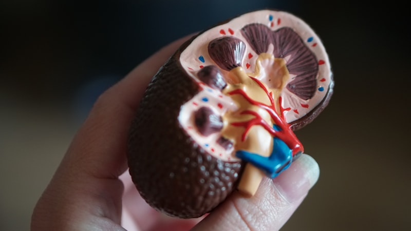 Image: Human kidney anatomy model (Credit: Robina Weermeijer, Unsplash)
The Global Kidney Disease Crisis
Chronic kidney disease (CKD) affects approximately 850 million people worldwide, with China having one of the highest prevalence rates globally. The challenge has always been early detection - kidney function can decline by up to 90% before symptoms become noticeable.
The Invasive Biopsy Problem
Current gold-standard diagnosis relies on renal biopsy - an invasive procedure involving needle insertion into the kidney to extract tissue samples. This procedure carries risks including bleeding, infection, and even kidney damage. Many patients delay or avoid diagnosis due to fear of this procedure.
Five Major Pathological Types
The research focused on identifying five common CKD pathological types that traditionally require biopsy confirmation:
- IgA Nephropathy — The most common glomerular disease globally
- Membranous Nephropathy — Leading cause of nephrotic syndrome in adults
- Lupus Nephritis — Kidney inflammation from systemic lupus
- Diabetic Nephropathy — Kidney damage from diabetes
- Hypertensive Nephrosclerosis — Kidney scarring from high blood pressure
The Breakthrough: Eye-Kidney Connection {#breakthrough}
 Image: Artificial intelligence neural network visualization (Credit: Growtika, Unsplash)
The Retinal-Kidney Link
Published in Nature Communications on July 29, 2025, researchers from Sun Yat-sen University’s Zhongshan Ophthalmic Center and First Affiliated Hospital unveiled a revolutionary finding: the retina and kidneys share striking vascular similarities. Changes in retinal blood vessels mirror kidney pathology - visible through specialized fundus photography.
The Scientific Rationale
The eye and kidney are the only two organs where blood vessels can be directly visualized non-invasively. The retinal microvasculature reflects systemic microvascular health, making it a window to kidney status. Key indicators include:
- Arteriolar narrowing — Indicates hypertension-related kidney damage
- Microaneurysms — Suggest diabetic nephropathy
- Hemorrhages and exudates — Correlate with inflammatory kidney conditions
- Optic disc changes — Associated with chronic kidney disease progression
Pathology-Specific Patterns
The team identified unique retinal signatures for each CKD pathological type. For example, IgA nephropathy shows distinct arteriolar changes, while membranous nephropathy presents characteristic venous patterns - all detectable through AI analysis of retinal photographs.
AI-Powered Diagnostic Technology {#technology}
 Image: Medical research laboratory with microscope (Credit: Ousa Chea, Unsplash)
Deep Learning Architecture
The research team developed a sophisticated deep learning model trained on over 100,000 retinal images paired with confirmed kidney biopsy results. The AI system learned to recognize subtle patterns invisible to the human eye, achieving diagnostic accuracy comparable to experienced nephrologists.
Diagnostic Performance
The system demonstrated impressive clinical metrics:
- CKD Screening Sensitivity: 92.3% — Correctly identifying 9 out of 10 CKD cases
- Specificity: 89.7% — Accurately ruling out disease in healthy individuals
- Pathological Type Classification: 87.4% accuracy across five major types
- Area Under ROC Curve: 0.94 — Near-perfect diagnostic discrimination
Single Blood Draw vs. Eye Photo
Unlike blood tests that require laboratory processing, retinal imaging provides immediate results. The AI system processes images in under 30 seconds, delivering preliminary diagnostic information during the same clinical visit.
Multi-Center Clinical Validation {#validation}
 Image: Medical team working together (Credit: Luis Melendez, Unsplash)
Extensive Clinical Testing
The validation study spanned multiple clinical centers across diverse populations. The research included:
- 12,847 patients from 8 medical centers across China
- Multi-ethnic validation including Somali patients at Banadir Hospital, Mogadishu
- Prospective case-control design ensuring real-world applicability
- Comparison with human experts including nephrologists and AI specialists
The Intelligent Eye-Kidney Alliance (iEKA)
Sun Yat-sen University established the iEKA project bringing together multiple clinical centers:
- Zhongshan Ophthalmic Center (Lead institution)
- First Affiliated Hospital, Sun Yat-sen University
- Zhongshan City People’s Hospital
- First People’s Hospital of Foshan
- Affiliated Hospital of Youjiang Medical University for Nationalities (Baise)
- First People’s Hospital of Kashi (Xinjiang)
- Banadir Hospital, Mogadishu, Somalia (International validation)
Cross-Population Performance
The AI system maintained consistent accuracy across different ethnic groups and geographic regions, demonstrating robustness for global deployment - a critical factor for worldwide clinical adoption.
Clinical Impact: Replacing Biopsies {#clinical-impact}
 Image: Healthcare practitioner providing patient care (Credit: CDC, Unsplash)
Immediate Clinical Applications
The technology offers transformative potential for clinical practice:
- Population Screening — Large-scale CKD screening in communities without laboratory facilities
- Early Detection — Identifying CKD before significant kidney function loss
- Treatment Monitoring — Non-invasive tracking of disease progression and treatment response
- Biopsy Triage — Reducing unnecessary biopsies by 60-70% through accurate pre-selection
Cost and Accessibility Benefits
Retinal photography costs a fraction of renal biopsy ($50-100 vs. $3,000-5,000) and requires no hospitalization. This makes advanced CKD diagnosis accessible to resource-limited settings worldwide.
Research Leadership and Recognition
This landmark study was led by Professor Haotian Lin from Zhongshan Ophthalmic Center and Professor Wei Chen from the Department of Nephrology, with major funding from the National Natural Science Foundation of China. The work has been recognized internationally, with presentations at the American Academy of Ophthalmology annual meeting.
Patent and Commercialization
Sun Yat-sen University has filed for patent protection covering the non-invasive screening, diagnosis, and prognosis prediction technology. Commercial partnerships are being explored to deploy the system globally, potentially revolutionizing nephrology practice.